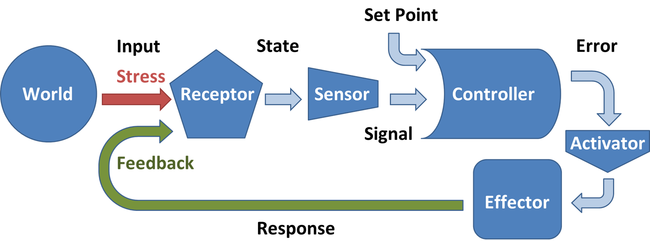
- Regulation of the water potential of the blood -
Osmoreceptors in the hypothalamus monitors the water potential in the blood.
If the water potential decreases in the blood, water would move out of the osmoreceptor cells by osmosis // cells decrease in volume as a result of this // signal is sent to other cells in the hypothalamus // signal then sent to the posterior pituitary gland. - this secretes the antidiuretic hormone (ADH) into the blood.
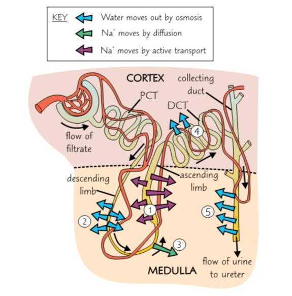
ADH makes the walls of the distal convoluted tubule and collecting duct more permeable to water.
More water is reabsorbed from these tubules into the medulla and into the blood by osmosis // small concentration of urine is produced = reduction of water loss.
More water is reabsorbed from these tubules into the medulla and into the blood by osmosis // small concentration of urine is produced = reduction of water loss.
Specific protein receptors on cell surface membrane of these cells binds to ADH molecules // activation of phosphorylase (enzyme) within cell // vesicles within the cell move and fuse with the cell surface membrane.
Vesicles contain plasma membrane and have numerous water channel proteins (aquaporins) so when they fuse with the membrane the number of water channels is considerably increased - making cell-surface membrane more permeable to water.
ADH increases permeability of collecting ducts to urea // passes out // lowering water potential of fluid around the duct.
Combining effect is more water leaves the collecting duct urea // osmosis // down a concentration gradient // re-enters the blood.
Reabsorbed water came from the blood will prevent the water potential lowering // Osmorecpetors send nerve impulses to the thirst centre of the brain encouraging the individual to seek out and drink more water.
Hypothalamus detects the rise in water potential and send fewer impulse to the pituitary gland
Pituitary gland reduces the release of ADH // permeability of the ducts return to its former state // Homeostasis and Negative Feedback.
A fall in the solute concentration of blood raises its water potential. Due to either:
1) Large volumes of water being consumed.
2) Salts used in metabolic or excreted not being replaced in the diet.
The body responds to rise in water potential:
Osmoreceptors in hypothalamus detects rise in water potential // increase frequency of the of nerve impulses to the pituitary gland to reduce its release of ADH
Less ADH // via blood // decrease in permeability of the collecting duct to water and urea
Less water is re-absorbed into the blood from the collecting duct ⇒ more dilute urine = water potential falls in the blood.
In the blood the water potential has returned to normal // osmoreceptors cause the pituitary gland to raise ADH // the levels go back to normal ⇒ Negative Feedback.