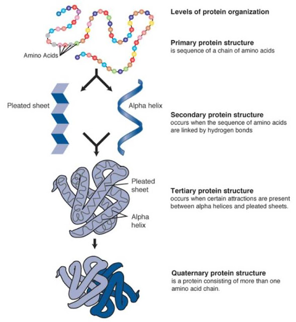
Showing posts with label Biological Molecules. Show all posts

Sunday, 23 April 2017

Biological Molecules: - Enzyme Inhibition

- Enzyme Inhibition - 


Enzyme inhibition: enzyme inhibitors are substances that directly or indirectly interfere with the function of the active site of an enzyme, reducing its activity.
Competitive inhibitors: inhibitors have a molecular shape which would be similar to the substrate, which allows them to occupy the active site instead of the substrate, competing with the substrate molecule.
If the substrate concentration is increased, the effect of the inhibitor rescued.
The inhibitors is not permanently bound to the active site thus when it leaves, another molecule can take it place.
The concentration of the inhibitor determines how long it will take for all substrate molecules to occupy the active site.
Image result for competitive inhibition


- Non competitive inhibitors -
They alter the shape of the enzyme and active site which means that substrate molecules can no longer occupy it and so the enzyme cannot function. Because the inhibitor and the substrate are not competing for the same site, an increase in substrate concentration does not decrease the effect of the inhibitor.


Image result for non-competitive inhibition

Biological Molecules: - Factors Affecting Enzyme Action

- Factors Affecting Enzyme Action - 

There are many factors that effect enzyme action, this includes:

  1. Temperature.  
  2. pH levels.
  3. Concentration
However before delving into detail about how they effect the enzyme action. First, we have to see how enzyme-catalysed reactions are measured. 


To measure the progress of an enzyme-catalysed reaction the time course is measured

2 changes that are frequently measured:

> Formation of the products of the reaction
> Disappearance of substrate.


Measuring rate of change 


The change in the rate of reaction can be measured at any point on the curve of a graph. 

The gradient is equal to the gradient of the tangent to the curve at the point the tangent is the point at which a straight line touches the curve but without cutting across it. 


You can draw the tangent to the curve at the point shown. Using this line you can find the gradient 

     a 
 = ㄧ     
     b

Effects of different factors on the rate of enzyme action it is important to stress the fundamental experimental technique of changing only a single variable in each experiment. 

Rate of an enzyme reaction all the other variables must be kept constant when investigating the effects of a named variable. 


Another thing to remember is that the active site and substrate is not the same as a lock and key.  

Temperature ● pH ● Concentration 
Factors effecting enzyme action. 


Image result for Factors Affecting Enzyme Action


Temperature: 

When there a temperature increase, there is an increase in kinetic energy of molecule. 
The molecules move around rapidly colliding with each other more often. 
Enzyme-catalysed reaction = enzyme and substrates molecules come together more often in a given time. 
More effective collisions = More enzyme-substrate complexes being formed

At first the substrate does not fit as easily into the changed active site = slowing rate of reaction. 
Human enzymes begin at temperature of 45℃. 
Around 60℃ enzyme is so disrupted its stops working together = the enzyme denatures
Denaturation is a permanent change and enzyme does not function again. 
Many enzymes have an optimum temperature of around 40℃ in the human body. 

Optimum temperature = the most/best favourable point or degree or condition at which an organism can work at which would obtain the best results


pH: 

pH of a solution is the measure of hydrogen ion concentration. 

The pH of a solution is calculated using : 

pH = -log₁₀[H⁺] 

H⁺ concentration of 1 x 10⁻⁹ therefore has a pH of 9 

An increase or decrease in pH reduces rate of enzyme action. If the pH level is extreme the enzyme becomes denatured. 

The pH level affects enzymes works in two ways:
  1. pH alters the charge on the amino acids that make up the active site of the enzyme = substrates can no longer attach to the active site. In other words, enzyme-substrate complex cannot be formed. 
  2. pH may cause the bonds maintaining the enzyme's tertiary structure to break = active site changes. 


Concentration: 

Image result for effect of enzyme concentration on rate of enzyme action

The active site can be reused to repeat the procedure on another substrate molecule once it is free. This means that enzymes are not used up during the chemical reactions, resulting in them working efficiently at very low concentrations.
  1. Low enzyme concentration: there are too few enzyme molecules to find an active site at one time.
  2. Intermediate enzymes concentration: all the substrate molecules can occupy an active site as the same time. The rate of reaction had doubled to it's maximum as a result.
  3. High enzyme concentration: adding more enzyme molecules has no effect as there are already enough active sites to accommodate all the substrate molecules.


Biological Molecule: - Non- Reducing Sugar Test

- Test For Non-Reducing Sugars - 


Test for non-reducing sugars: some disaccharides are reducing sugars and we can use Benedict's test to detect them. Sucrose is known as a reducing sugar because it doesn't change the colour of Benedict's reagent when heated with it.


  1. Liquify sample.
  2. Add 2cm³ of the food sample to 2cm³ of Benedict's reagent.
  3. Place the test tube in a gently boiling water bath for 5 minutes. If the Benedict's reagent does not change colour then a reducing sugar is not present.
  4. Add another 2cm³ of the food sample to 2cm³ of dilute hydrochloric acid in a test tube and place the test tube in gently boiling water bath for 5 minutes. The hydrochloric acid will hydrolyse any disaccharides present into its constituent monosaccharides.
  5. Add sodium hydrogen carbonate solution to neutralise the hydrochloric acid. Test with pH paper to check that solution is alkaline
  6. Retest the resulting solution, if non reducing sugar present the sample would turn orange-brown colour


Image result for test for reducing sugars

Biological Molecule: - Reducing Sugars Test

- Test For Reducing Sugars - 


Test for reducing sugars:
All monosaccharides and some disaccharides are reducing sugars. A reduction reaction is a chemical reaction involving the gain of electrons or hydrogen. Reducing sugars that can donate electrons to another chemical. Reducing sugars can be tested for using Benedict's reagent which is an alkaline solution of copper II sulphate. An insoluble red precipitate of copper oxide is formed when reducing sugars are heated with benedict's reagent.
Methods:

  1. Add 2cm³ of the food sample to be tested to be tested to the test tube (this should be in liquid form).
  2. Add an equal volume of Benedict's reagent.
  3. Heat the mixture in a gently boiling water for 5 minutes.

Image result for carbohydrates structure

Biological Molecules: - Test For Lipids

- Test For Lipids - 


Test for lipids:
  1. Take a completely dry and grease-free test tube.
  2. Add 5cm³ of ethanol.
  3. Shake the tube thoroughly to dissolve any lipid in the sample.
  4. Add 5cm³ of water.
  5. A cloudy - white colour indicates indicates the presence of a lipid.
  6. Repeat using water instead of the sample, the final solution should remain clear.
The cloudy colour is due to any lipid in the sample being finely dispersed in the water to form an emulsion.

Image result for test for lipids

Biological Molecules: - Tests for Starch

- Test For Starch - 

Test for starch:


  1. Place 2cm³ of sample being tested into a test tube.
  2. Add a few drops of iodine solution and shake or stir.
  3. The presence of starch is indicated by the blue-black coloration.


Image result for test for starch in food

Monday, 17 April 2017

Biological Molecules: - Enzyme Action

- Enzyme Action -


Enzymes are important in everyday life, without us realising it. They assist us in respiration, metabolic reactions, digestion, etc.. They are an important in serving a wide range of important functions in the body. 


Enzymes = Globular proteins // Acts as catalysts. 

So how do enzymes as a, catalysts, lower the activation energy?


Image result for how enzymes lower activation energy


For enzymes to assist in a chemical reaction there would need to be activation energy. 

Activation energy = the minimum quantity of energy which the species must have in order to undergo with the specific reaction

Enzymes allow reactions to occur at lower temperatures than normal -  this is a result of the enzyme lowering the activation energy. 
For example - some metabolic processes occur at the human body temperature of 37℃ ⇒ this is relatively low for a chemical reaction. 
Without enzymes, reactions would proceed too slowly to sustain life. 


What is the structure of an enzyme? 


A specific region of the enzyme is classed as functional - active site

The active site is made up of small number of amino acids. Within the much large enzyme the active site would leave a small depression to allow the substrate to fit

The substrate is the molecule which the enzyme acts on. When the substrate fits into the depression it forms an enzyme-substrate complex. This is held by bonds that is temporarily formed between the certain amino acids on the active site and groups on the substrate molecule. 


Induced fit model of enzyme action. 


Image result for enzyme structure a level


Induced fit model is a more recent model developed, that is both widely accepted and has evidence to back it up.

The model states that the active site of the enzyme is not exactly complementary to the substrate. However, in the presence of a specific substrate the shape of the active site would change slightly in order to become complementary. 

In other words, the enzyme is flexible and is able to mould itself around the substrate like a glove moulds itself around a hand. 

The Lock and Key model - 

This was a much earlier model explaining the enzyme action. Suggesting enzyme action was the same as a lock and key. Each lock has a specific key, the key would only operate and unlock the specific lock. 
This implies that the substrate shape is specific to the active site and has an exact fit. 

A limitation of this would be that the model considers the enzyme as a rigid structure, but as it was found other molecules could bind to the enzyme at sites that was not the active site. As a result of this the enzyme structure was being altered. 

The structure was not rigid but flexible. 


http://alevelnotes.com/content_images/lock-and-key_model.jpg


Sunday, 16 April 2017

Biological Molecules: - Protein

- Protein - 


Structure of an amino acid

Image result for amino acids protein

Amino acid is the back bone monomer units 

Polymer ⇒ Polypeptide

Polypeptides ⇒ proteins

Every amino acid has a central carbon attached four different chemical groups

>  Amino group -NH2 - which the amino part of the name of the amino acid is derived. 

> Carboxyl group -COOH - acidic group gives the amino acid the acid part of the name. 

> Hydrogen atom - H. 

>R (side) group - variety of different chemical group 20 naturally occurring amino acids differ only in their R (side) group. 


Image result for amino acid structure


The formation of a peptide bond 


A peptide bond formed through a condensation reaction as a result of joining two or more amino acids together. 
The water is formed by combining an -OH from the carboxyl group of one of the amino acid with the -H of the other amino acid.
The amino acids are linked by a peptide bond between the carbon atom of one amino acid and the nitrogen atom of the other. 

There are four structures of the protein molecule.

- Primary structure 

- Secondary structure  

- Tertiary structure 

- Quaternary structure 

Image result for primary structure of protein


Primary structure

Polymerisation ⇒ This is a series of condensation reactions of many monomers of amino acids. As a result of this there is a long chain of amino acids (Polypeptide) 

A sequence of amino acids in a polypeptide is the back bone formation of the primary structure of any protein, which is determined by DNA. 

There are 20 naturally occurring amino acids joined in different sequences
The primary structure determines the ultimate shape and function, a single change made to the amino acid sequence can lead to a change in shape of the protein and may stop its function completely. 

Proteins have a specific shape which is specific to its function


Image result for primary structure of protein

Secondary structure 

The polypeptide chain is twisted into a 3-D shape ( coiled - α-helix ) 
This is due to the -NH and the -C=O the -NH has a hydrogen with a overall positive charge whereas the -C=O has an overall negative charge. 
This readily forms weak hydrogen bonds


Image result for secondary structure of protein

Tertiary structure 

The α helices of the secondary protein structure is twisted & folded to form an even more complex, 3-D and specific structure

There are 3 bonds that maintains this Polypeptide helix structure. 

- Disulfide bridges = fairly strong, not easily broken
- Ionic bond = formed between carboxyl and amino group // not involved in formation of peptide bonds. Not as strong as Disulfide bonds // Easily broken - by change in pH. 
- Hydrogen bonds = many of them // easily broken 


Image result for tertiary structure of protein


Quaternary structure 

A number of individual polypeptides chains are linked together in various ways. 
This structure, sometimes, includes non-protein // prosthetic// group i.e. iron containing haem group in haemoglobin. 


Image result for quaternary structure of protein


--------------------------------------------------------------------------------------------------------------------------

There are two types of protein: 

Fibrous protein and Globular protein 

If you think of polypeptide as a string then you can understand that: 

Fibrous protein is like collective pieces of string being twisted to for rope, 
whereas, 
Globular protein is like fewer pieces of string that is being rolled into a ball. 

--------------------------------------------------------------------------------------------------------------------------


Biological Molecules: - Lipids

- Lipids - 


- The Role Of Lipids - 


S ● W ● I ● P 


This helped me remembering the roles of lipids -

S ⇒ Source of energy 

lipids provide twice the energy as carbohydrates at the same mass when hydrolysed

W ⇒ Waterproofing 

Lipids are insoluble in water // plants and insects have a waxy cuticle reducing water loss whereas mammals have an oily secretion from sebaceous gland in the skin. 

I ⇒ Insulation 

Fats = slow conductor of heat // stored beneath the body surface to help retain heat 

P ⇒ Protection 

Fat stored around delicate organs i.e. Kidneys.

There are 2 types of lipids 

Phospholipids and Triglyceride 

So what is the difference?

Phospholipids have two parts

To remember there are two parts 

PHOSPHOLIPIDS = PHO is repeated twice within the word - therefore two parts :) 

Triglycerides have 3 fatty acids. 

To remember there are 3 fatty acids.

TRIGLYCERIDE = TRI means 3 


- Phospholipids -

- The two parts of the phospholipids is:

Hydrophilic 'head' = interacts with water (attracted to) // does not readily mix with fat. 

Hydrophobic 'tail' = orients itself away from water (repels against) // readily mixes with fat. 

- It has two ends and is said to be polar as they behave differently

- Structure of phospholipids 

⇒ Phospholipids are polar molecules - in aqueous environment phospholipid bilayer is formed hydrophobic barrier is formed between the cell (inside and outside).

⇒ Hyrdophilic 'head' helps hold at the surface of the cell-surface membrane.

⇒ Structure allows the formation of glycolipids // combining carbohydrates with the cell surface membrane. Acts as a cell recognition site.


top phosphate which is hydrophillic. middle is lipid which is hydrophobic bottom phosphate.  all human cells have this phospholipid bilayer surrounding their cell.  n.b transporters, channels and receptors


- Triglycerides - 

3 fatty acids joined to a glycerol 

( TRI = 3 // GLYCERIDE = glycerol. )

The bond formed = ester bond (through condensation reaction

There are 70 different types fatty acids, all have a carboxyl ( -COOH ) group attached with a hydrocarbon chain. 

If this chain does not have carbon-carbon double bond = Saturated 

If this chain has a single double bond = Mono-UnSaturated 

If this chain has more than one double bond = Polyunsaturated

To remember the difference between unsaturated and saturated. 

--------------------------------------------------------------------------------------------------------------------------

UN-SATURATED = There are two parts to the word which can imply double bond 

SATURATED = A single word which can imply a single bond. 

--------------------------------------------------------------------------------------------------------------------------

Structure of triglycerides - 

⇒ High ratio of energy-storing carbon-hydrogen bonds to carbon atoms // excellent source of energy. 

⇒ Low mass to energy ratio // good storage molecule = so much energy can be stored in small volume. Reduced mass in animals so able to move around. 

⇒ Large, non polar & insoluble // storage does not effect osmosis in cells or the water potential 

⇒ High ratio of hydrogen to oxygen atoms // release water when oxidised provides an important source of water. 

Image result for triglycerides


Biological Molecules: - Carbohydrates


- Carbohydrates - 


- Monosaccharides - 


The monomer for carbohydrates is called monosaccharides. 

Common forms of monosaccharides are 
- Glucose 
- Galactose 
- Fructose 

In order for a two monosaccharides to form, there needs to a condensation reaction.

The bond that is formed between = glycosidic bond.

2 monosaccharides with a glycosidic bond in between = Disaccharide


- Disaccharides -


Examples of this = 


Image result for disaccharides


The picture above shows the different types of disaccharides and the structures for each of them. 


Glucose is needed for each of the disaccharide formation. 

This is because Glucose is an isomer

Glucose has 2 isomers - 

- (Alpha) α-glucose 

- (Beta) β-glucose 


Image result for alpha and beta glucose


As shown above which has been circled, the positioning of the OH is the difference in the structure of both molecules. This can make all the difference in a molecule, which can change its function, structure, etc. 


- Polysaccharides -

A polysaccharide is formed from many glucose units by condensation. 

There are 3 forms of polysaccharides that are needed to be known: 

- Glycogen } formed by the condensation of α- glucose.

- Starch } formed by the condensation of α- glucose.

- Cellulose } formed by the condensation of β- glucose. 


Glycogen vs Starch 

Starch: 

Main role = energy store 
Structure is suited for the molecule, reasons being it's:
 Insoluble ⇒ does not effect water potential (water not drawn by osmosis).
large & insoluble ⇒ does not leave the cell.
compact ⇒ can be stored in small places.
hydrolysedα- glucose, can be transported & readily used in respiration.
branched form = enzymes, simultaneously, can act on the polymer releasing α- glucose.
Glycogen: 
Main role = major carbohydrate store in animals
 Insoluble = does not effect water potential (osmosis) // does not diffuse out of the cell.  
 Compact = huge amounts can be stored in a small space
 Highly branched = ends are acted on simultaneously by enzymes, rapidly broken to form glucose monomers - respiration. 


Image result for glycogen vs starch


Cellulose

Unlike Starch and Glycogen, cellulose is not formed from α- glucose. 

β- glucose is building blocks of cellulose. The small variation produces a fundamental difference in the structure and the function of the polysaccharide. 

Cellulose:
Main role = providing support and rigidity
forms long single un-branched chains.
run parallel to each other // crossed linked by hydrogen bonds add collective strength. 
 grouped to form micro-fibrils which are grouped forming fibres // adds more strength. 


Image result for cellulose

Biological Molecules: -


Monomers and Polymers

I must admit I did skip this section when revising last year and to my fault of understanding this was the foundation for my learning.


I made this mistake so you guys don't!
--------------------------------------------------------------------------------------------------------------------------
So what is a monomer?

"A monomer is smaller units from which larger molecules are made." 
Monomers are important in the formation of other polymers. For example,

Glucose + Glucose = Maltose

This an example of monosaccharides. Other examples of monomers =
- amino acids
- nucleotides.

So what is a polymer?

"A polymer is molecules made from a large number of monomers joined together." 
In order for the polymers to be formed there would need to be bonds between the molecules to hold the structure together, right?


Image result for Monomers  ← this shows the structure of monomers and polymers which is always useful to know :) 


How do monomers lead to the formation of polymers?


When monomers join together, a condensation reaction occurs which forms a chemical bond and involves the elimination of a water molecule. Thus forming a polymer!


Compound Interest


How do polymers break down to then form monomers?

A hydrolysis reaction!!

This is when the chemical bond between two molecules is broken through the use of a water molecule.


Compound Interest

Biological Molecules: - Carbohydrates

- Carbohydrates -   - Monosaccharides -  The monomer for carbohydrates is called monosaccharides.  Common for...