Sunday, 30 April 2017

Organisms Respond To Changes In Their Environment: - Role Of The Nephron In Osmoregulation

- Nephron filtering the blood -


  1. Blood from the renal artery enters smaller arterioles in the cortex of the kidney.
  2. Arterioles splits into a glomerulus // bundle of capillaries looped inside a Bowman's capsule // Ultrafiltration takes place.
  3. Efferent arterioles that takes blood into each glomerulus // afferent arteriole take the filtered blood away from the glomerulus
  4. Efferent arterioles are smaller in diameter than afferent arterioles // blood is under high pressure. 
  5. High pressure forces liquid and small molecules in the blood out of the capillary and into the Bowman's capsule
  6. Liquid and small molecules pass through 3 layers get into Bowman's capsule enter the nephron tubules - capillary wall // basement membrane and epithelium of Bowman's capsule.
  7. Larger molecule - protein and blood cells can't pass through - stay in blood. Substances enter Bowman's capsule // glomerular filtrate 
  8. Glomerular filtrate passes along the rest of nephron - useful substances are reabsorbed along the way
  9. Filtrate flow through the collecting duct and passes out of the kidney along the ureter


Re-absorption of glucose and water by the proximal convoluted tubule
The proximal convoluted tubule is adapted to reabsorb substances: 
Its adaptations include:
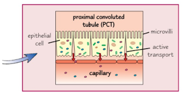
- micovilli = large surface area to reasorb substances from filtrate 
- infolding of bases = large surface area to transfer reabsorbed substances back into the blood capillaries 
- high density of mitochondira to provide ATP // active transport

Process of re-absorbtion 

  1. Sodium ions actively transported out of the cells lining proximal convoluted tubule into blood capillaries // sodium concentration drops 
  2. Sodium ions diffuse down a concentration gradient from lumen of proximal convoluted tubule into epithelial lining cells // only through carrier proteins // facilitated diffusion. 
  3. Carrier proteins are specific types to carrier other specific molecules// co-transport // molecules which have been co-transported into the cell of proximal convoluted tubule then diffuse into the blood // All glucose and other valuable minerals are reabsorbed 
Maintenance of gradient of sodium ions by the loop of Henle 

Loop of Henle = responsible for water being reabsorbed from the collecting duct // concentrating the urine so it has lower water potential than blood. 

This has 2 regions 
  1. Descending limb - narrow - thin walls that are highly permeable to water. 
  2. Ascending limb - wider - thick walls that impermeable to water.

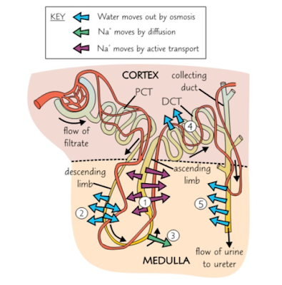
  1. Na+ ions are pumped out into the medulla using active transport // ascending limb impermeable therefore water does not move out lowering the water potential in the medulla, because there's a high concentration of ions
  2. Due to there being a lower water potential in the medulla than in descending limb, water moves out the descending limb, into medulla by osmosis // filtrate is more concentrated. The water in the medulla is reabsorbed into the blood through the capillary network
  3. Bottom of the ascending limb // Na+ ions diffuse out into the medulla, lowering the water potential further in the medulla. The ascending limb is impermeable to water, so it stays in the tubule. 
  4. Water moves out of distal convoluted tubule by osmosis and is reabsorbed into the blood.
  5. First three stages massively increase the ion concentration in the medulla, lowers the water potential. Water then moves out of the collecting duct by osmosis. The water in the medulla is reabsorbed into the blood through the capillary network


Organisms Respond To Changes In Their Environment: - Control of Blood Water Potential


- Structure Of The Nephron - 


Osmoregulation = the homeostatic control of the water potential of the blood

Structure of the mammalian kidney

In the mammal there are two kidneys found at the back of the abdominal cavity
A section through the kidney 

Image result for structure of mammalian kidney showing the position of two of the million or more nephrons in each kidney

Fibrous Capsule = outer membrane that protects the kidney 

Cortex = lighter coloured outer region made up of renal (Bowman's) capsule, convoluted tubles and blood vessels. 

Medulla = darker coloured inner region made up of loops of Henle, collecting ducts and blood vessels 

Renal pelvis = funnel-shaped cavity that collects urine into the ureter 

Ureter = tube that carries urine to the bladder 

Rental artery = supplies the kidney with blood from the heart via the aorta

Renal vein = returns blood to the heart via the vena cava 


Structure of the nephrons 

Renal (Bowman's) capsule = closed end at the start of the nephron. It cup-shaped and surrounds a mass of blood capillaries - Glomerulus. The inner layer of the renal capsule made of specialised cells - podocytes 

Proximal convoluted tubules = series of loops surrounded by blood capillaries. It's walls made epithelial cells - have microvilli 

Loop of Henle = long hairpin loop extends from cortex into medulla of kidney and back again. Surrounded by blood capillaries 

Distal convoluted tubule = series of loops surrounded by blood capillaries. Walls are made of epithelial cell - surrounded by fewer capillaries than proximal tubule

Collecting duct = tube number of distal convoluted tubules from a number of nephrons empty. Lined by epithelial cells and becomes increasingly wide as it empties into the pelvis of kidney


Associated with each nephron there are number of blood vessels:

Afferent arteriole = tiny vessel arise from renal artery - supplies the nephron with blood // Afferent arteriole enters the renal capsule of the nephron 

Glomerulus = many-branched knots of capillaries from which fluid is forced out of the blood. Glomerulus capillaries recombine.

Efferent arterioles = tiny vessel leaves the renal capsule // smaller diameter than afferent arteriole causes an increase in blood pressure within the glomerulus. Efferent arterioles carries blood away from the renal capsule

Blood capillaries = concentrated network of capillaries surrounding the proximal convoluted tublue, loop of Henle mineral salts, glucose, and water. These capillaries merge to form venules, merging to form the renal vein


Organisms Respond To Changes In Their Environment: - Diabetes And Its Control

- Diabetes and its control - 


Types of sugar diabetes 


Type 1 - insulin dependent // body unable to produce insulin 

The body attacks its own cells - 𝛽 cells - result of autoimmune response. 
This develops quickly over a few weeks once the individual is born. Usually develops at a very young age. 

Type 2 - insulin independent // body glycoproteins receptors have lost or losing their responsiveness to insulin // body producing an insufficient amount of insulin
This develops in people over age of 40 - increases due to obesity and poor diet
Symptoms are less severe and may go unnoticed
90% of people have type II diabetes 


Control of diabetes 

Diabetes can be successfully treated which depends on the type of diabetes 

Type 1 diabetes = controlled by injections of insulin - cannot be taken orally as it is an enzyme and would be digested. Typcially taken 3-4 times a day. Dose of insulin would match the dose intake of glucose // glucose concentration is monitored using biosensors

Type 2 diabetes = controlled by regulating the intake of carbohydrates in the diet // exercise is also taken into account. Supplemented by injections of insulin by the use of drugs that stimulate insulin production


Image result for insulin type 1 and type 2


Organisms Respond To Changes In Their Environments: - Hormones And The Regulation Of Blood Glucose Concentration

- Hormones and their mode of action - 

Hormones differ chemically but there are characteristics which would be the same also. 

Hormones :

- Produced in glands // secrete hormones directly into blood
- Carried in blood plasma to cells which are acted on // target cells // specific receptors on their cell-surface membrane // complementary to specific hormones.
- Effective in very low concentrations // widespread // long-lasting.


 Second Messenger Model 

2 hormones are used - Adrenaline and glucagon 

  1. Adrealine binds to the transmembrane protein receptor within the cell-surface membrane of the liver cell. 
  2. Binding of the adrealine causes the protein to change the shape on the inside of the membrane
  3. Change in shape of the protein leading to the activation of an enzyme - adenly cyclase // activated adenly cyclase converts ATP to cyclic AMP (cAMP) 
  4. cAMP acts as second messenger that binds to the protein kinase enzyme, its activiated by changing its shape.
  5. Active protein kinase enzyme catalyses the conversion of glycogen to glucose which moves out of the liver cell // facilitated diffusion // in to the blood through channel proteins
Image result for second messenger model

Role of the Pancrease in regulating blood glucose 

It produces enzymes - 
Protease, amylase and lipase for digestion and hormones
Insulin and glucagon for regulating blood glucose concentration

There are groups of hormone-producing cells - islets of Lnagerhans - cells of the islets of Langerhans:
- ɑ cells, larger and produce the hormone glucagon
- 𝛽 cells, smaller and produce the hormone insulin 

Role of the liver in regulating blood sugar 

There are three important processes involved with regulating blood sugar which takes place in the liver:

Glycogenesis - this is the conversion of glucose into glycogen
This occurs when the blood glucose concentration is higher than normal. The liver removes the glucose from the blood and converts it into glycogen

Glycogenolysis - this is the breakdown of glucagon into glucose.
This occurs when the blood glucose concentration is lower than normal. The liver converts glycogen into glucose which is then diffused into the blood

Gluconeogenesis - this is production of glucose from sources other than carbohydrates
This occurs when glycogen supply is exhausted. The liver produces glucose from non-carbohydrate sources - amino acids and glycerol


Regulation of blood glucose concentration 


Factors that influence blood glucose concentration
  1. Directly from the diet = glucose is absorbed from the hydrolysis of carbohydrates 
  2. From the hydrolysis of glycagon in the small intestine = glycogenolysis // stored in liver and muscle cells 
  3. From gluconeogenesis 


Insulin and the 𝛽 cells of the pancreas 
when combined with receptors insulin brings about:

- Change in tertiary strucute of the glucose transport carrier proteins - changing shape and open - allows more glucose into the cells by facilitated diffusion.
- Increase in number of carrier proteins responsible for glucose transport in the cell-surface membrane. Protein from which these channels are made is part of the vesicles. // rise in insulin concentration the vesicles fusing with the cell-surface membrane increasing the number of glucose transport channels. 
- Activation of enzyme that convert glucose to glycogen and fat

Blood glucose is lowers through the following ways:

- Increasing the rate of absorption of glucose into the cells - muscle cells
- Increasing rate of respiration of the cells // uses more glucose // increasing the uptake from the blood
- Increasing glycogenesis in liver and muscle cells
- Increasing the rate of conversion of glucose to fat

The processes taking place = Negative feedback // removal of glucose reduced the secretion of insulin from 𝛽 cells


Glucagon and the ɑ cells of the pancreas 
ɑ cells detect the fall in blood glucose concentrations releasing the hormone glycogen directly into the blood plasma
Glycogen action include: 
- Attaching to specific protein receptors on cell-surface membrane of liver cells 
- Activating enzymes convert glycogen to glucose 
- Activating enzymes involved in conversion of amino acids and glycerol into glucose = glyconeogenesis 
This returns the blood glucose concentration to the optimum

Image result for alpha and beta cells in the pancreas

Role of adrenaline in regulating the blood glucose level 

Adrenaline raises the blood glucose concentration by:
- Attaching to protein receptors on the cell-surface membrane of target cells 
- Activating enzymes that causes the breakdown of glycogen to glucose in the liver

Adrenaline is produced when there is a period of excitement or stress

Hormone interaction in regulating the blood glucose

Insulin and glycagon act in opposite directions = antagonistically 
These two hormones are sensitive to control of the blood glucose concentration // blood glucose concentration is not constant there are fluctuations around the optimum temperature

When writing about negative feedback its important to mention that the secretion of a hormone - insulin - would result in the reduction of its own secretion


Organisms Respond To Changes In Their Environments: - Feedback Mechanisms

- The control mechanisms -

Control of any self regulating system involves a series of stages

- Optimum point
- Receptor
- Coordination
- Effector
- Feedback mechanism

Homeostatic systems detect change and respond by negative feedback.

1) The receptorcommunication system and effectors is the foundation of the homeostatic system
2) Receptors are there to detect when there is a change in temperature - this would be when temperature is too high or too low. Via the nervous system or hormonal system the information is passed on to the effector. 
3) The effectors are suppose to counteract the change // bring the level back to an optimum
4) Negative feedback = process of bring the level to an optimum temperature
5) Negative feedback keeps the body's temperature at an optimum which is above 0.5℃ or below 37℃.
6) However if they change is too drastic then the Negative feedback would be unable to counteract the change. This can be when there has been prolonged exposure to cold weather


There are multiple Negative feedback mechanisms ⇒ more control. By only having one negative feedback system ⇒ there is a slower response and less control

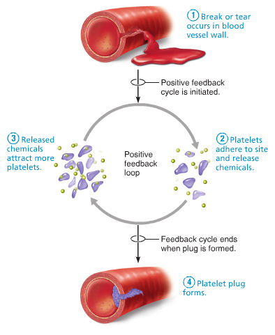
Positive feedback mechanisms can amplify a change from the normal level
1) some changes can trigger a positive feedback // amplifying the change. 
2) Increasing the level further away from the normal level.
3) Positive feedback rapidly activates something // Blood clot after a deep cut. 
4) Positive feedback can occur when homeostatic system breaks down e.g. Being too cold for too long.

Positive feedback is not involved in homeostasis because it does not keep the internal environment stable.


Image result for positive feedback homeostasis

- Coordination of control mechanisms -

Control system normally have receptors and effectors allows them to have separate mechanisms that produce a positive movement towards an optimum. Allows greater degree of control of the particular factor being regulated. 


Nucleic Acids: - Water

- Water - 

Dipolar water molecule: water is made up of two atoms of hydrogen and one of oxygen. The molecule has no overall charge although oxygen has a slightly negative charge and hydrogen has a slightly positive charge.
Water and hydrogen bonding:
A positive pole of one water molecule would attract the negative pole of another water molecule, the attraction force between the opposite charges is a hydrogen bond. Each bond is weak, however together they form important forces that cause water molecules to stick together, giving water unusual properties.

Image result for water structure
Specific heat capacity:

Water molecules stick together, it takes more energy -heat- to separate them then if they did not bond to one another. Resulting in the boiling point being high (100 degrees celsius)
Without hydrogen bonding water would be a gas at temperatures on earth.
Water acts as a buffer against sudden temperatures variations - aquatic environment is a temperature stable one.
Organisms are mostly water, protects them from sudden temperature change especially in terrestrial environment.
Latent heat of vaporisation of water:
Hydrogen bonding = lots of heat energy required to evaporate 1 gram of water. - latent heat of vaporisation.
Evaporation of water in mammals is therefore a very effective means of cooling, body heat is used to evaporate the water.
Cohesion and surface tension:
Tendency of molecules sticking together = cohesion
  • Hydrogen bonding = large cohesive forces, allowing it to be pulled through a tube - xylem vessel
  • Water molecules meet air, tend to be pulled back into the body of water than escaping from it = surface tension.
  • Water acts like a skin, strong enough to support small organisms - pond skaters

Importance of water to living organisms:
Water in metabolism -
  • Used to breakdown complex molecules - hydrolysis
  • Joins molecules together through the use of condensation reaction
  • Chemical reactions take place in aqueous medium
  • Water = major raw material in photosynthesis
Water as a solvent -
  • Readily dissolves other substances - gases ie, oxygen + carbon dioxide
  • Waste - ammonia + urea
  • Inorganic ions + small hydrophilic molecules - amino acids, monosaccharides, + ATP
  • Enzymes - reactions which takes place in a solution.

Important features of water:

  • Evaporation cools and allows organisms to control their temperature.
  • Not easily compressed = providing support - hydrostatic skeleton of animals// earthworm + turgor pressure in herbaceous plants
  • Transparent = aquatic plants can photosynthesise // light penetrate jelly like fluid that fills the eye and reach the retina.


Nucleic Acids: - ATP

- ATP - 


ATP - Adenosine triphosphate is a nucleotide and has three phosphate groups and is key in storing energy. The bonds between the phosphate groups are unstable therefore have a low activation energy - can be easily broken. They release a considerable amount of energy. In living cells it is only the terminal phosphate that is removed

Structure of ATP:
  • Adenine - nitrogen containing organic base.
  • Ribose - sugar molecule with 5 carbon ring structure - backbone
  • Phosphates - chain of three phosphate groups
.
Synthesis of ATP: ATP to ADP is a reversible reaction therefore energy can be used to add an inorganic phosphate to ADP to re-form ATP. This reaction is catalysed by enzyme ATP synthase. The reaction is a condensation reaction.

Image result for structure of ATP
The synthesis ATP from ADP involves addition of phosphate molecule to ADP.
  • In chlorophyll containing plant cells during photosynthesis (photophosphorylation)
  • In plant and animal cells during respiration (oxidative phosphorylation)
  • In plant and animal cells when phosphate groups are transferred from donor molecules to ADP (substrate level phosphorylation)
Roles of ATP:
  • ATP serves as an immediate energy source of a cell
  • Cells do not require large quantities of ATP
  • ATP is rapidly re-formed from ADP and inorganic phosphate
ATP is required as energy-requiring processes in cells:

  • Metabolic processes
  • Movement
  • Active transport
  • Secretion
  • Activation of molecules


Thursday, 27 April 2017

Organisms Respond To Changes In Their Environments: - Principles Of Homeostasis

- Principles Of Homeostasis -

What is Homeostasis?

This is the maintenance of an interval environment within restricted limits in organisms.
This is to ensure that the chemical make-up, volume and other features of blood and tissue fluid within restricted limits are maintained.

Homeostasis is the maintenance of a stable internal environment.
Any changes that are made to the external environment can affect the internal environment of an organism.

To prevent this, homeostasis is involved in control systems that would maintain the constant internal environment.
This allows virtual cells to function normally and prevent damage to them and they are kept in a stable environment.
Factors that would be maintained ⇒ the core body temperature and blood pH.

Image result for Homeostasis

This is because both pH and temperature are factors that effect enzyme activity and enzymes control the rate of metabolic reactions.

Temperature: if it is too high or at an extreme the enzymes would become denatured.
This is due to the hydrogen bonds breaking that holds them in their 3D shape. Thus changing the active site, which leads to an ineffective catalyst.
If the temperature is too low the enzyme activity is reduced and it slows down the rate of metabolic reactions.

In order for the rate of enzyme activity to remain at a constant the enzymes have an optimum temperature at which they work best at. In humans, this would be 37oC.

pH: if the pH is too high or too low // at either ends of extremes// the enzymes denature. Hydrogen bonds that hold the shape of the enzymes are broken. Therefore, the active site of the enzyme is changed which makes it ineffective catalyst. The metabolic reactions are less efficient.
The highest rate of enzyme activity remains at an optimum pH // usually remains around pH 7.
But this varies for some enzymes // enzymes for the stomach work best at a low pH.
Because cells need glucose for energy it is important to maintain the right concentration of glucose in the blood.

Image result for Homeostasis blood pH
Blood glucose concentration also affects the water potential of blood - there is the potential that water molecules diffuse in and out of or into a solution.
If blood glucose concentration is high ⇒ water potential of blood is reduced // water molecules would then diffuse out of the cell // osmosis // cell will shrivel up and die.

If blood glucose concentration is too low ⇒ cells cannot carry out normal activities there isn't enough glucose for respiration to provide energy.


- The control mechanisms -

Control of any self regulating system involves a series of stages

- Optimum point
- Receptor
- Coordination
- Effector
- Feedback mechanism

Homeostatic systems detect change and respond by negative feedback.

1) The receptor, communication system and effectors is the foundation of the homeostatic system
2) Receptors are there to detect when there is a change in temperature - this would be when temperature is too high or too low. Via the nervous system or hormonal system the information is passed on to the effector. 
3) The effectors are suppose to counteract the change // bring the level back to an optimum
4) Negative feedback = process of bring the level to an optimum temperature
5) Negative feedback keeps the body's temperature at an optimum which is above 0.5℃ or below 37℃.
6) However if they change is too drastic then the Negative feedback would be unable to counteract the change. This can be when there has been prolonged exposure to cold weather


There are multiple Negative feedback mechanisms ⇒ more control. By only having one negative feedback system ⇒ there is a slower response and less control

Positive feedback mechanisms can amplify a change from the normal level
1) some changes can trigger a positive feedback // amplifying the change. 
2) Increasing the level further away from the normal level.
3) Positive feedback rapidly activates something // Blood clot after a deep cut. 
4) Positive feedback can occur when homeostatic system breaks down e.g. Being too cold for too long.

Positive feedback is not involved in homeostasis because it does not keep the internal environment stable.


Image result for positive feedback homeostasis

- Coordination of control mechanisms -

Control system normally have receptors and effectors allows them to have separate mechanisms that produce a positive movement towards an optimum. Allows greater degree of control of the particular factor being regulated. 


Sunday, 23 April 2017

Nucleic Acids: - DNA Replicationn

- DNA Replication - 
DNA replication: cell division occurs in 2 main stages:
  • Nuclear division: nucleus divides
  • Cytokinesis - the whole cell divides
Semi-conservative replication ~ this ensures the genetic continuity between generations.

The enzyme DNA helicase breaks the hydrogen bonds between complementary bases in polynucleotides strands, causing the double helix to unwind/unravel.


.
The enzyme DNA polymerase joins adjacent nucleotide's in a condensation reaction.


The exposed nucleotide's act as a template to which the complementary free nucleotide's bid by specific base pairing


2 identical molecules of DNA are formed. This is due to each molecule retaining half of the original DNA materialWatson and Crick model of DNA replication suggested that the original DNA molecule splits into 2 separate strands therefore each of the new molecules would have one strand of the original material. This would be the semi-conservative model.  The conservative model suggests that the original molecule remained intact and the other molecule formed would be made of entirely new material

Image result for the conservative replication


Nucleic Acids: - Structure of RNA and DNA

- Structure of DNA and RNA -


Deoxyribonucleic acid and ribonucleic acid are important information-carrying molecules. In all living cells, DNA holds genetic information and RNA transfers genetic information from DNA to the ribosomes.
DNA molecule is a double helix with two polynucleotides chains held together by hydrogen bonds between specific complementary base pairs.
  • Adenine is complementary to thymine
  • Guanine is complementary to cytosine
Ribosomes are formed from RNA and protein.
Both DNA and RNA are polymers of nucleotides. Each nucleotides is formed from

  • Pentose sugar
  • Phosphate group
  • Nitrogen-containing organic base

Nitrogen containing bases are:
  • Adenine - A
  • Thymine - T
  • Guanine - G
  • Cytosine - C
  • Uracil - U ( RNA molecules only // replaces Thymine (T))
Pentose sugar, phosphate group and organic base are joined as result of condensation reactions, forming a mononucleotide.
2 mononucleotides may in turn be joined by the condensation reaction between the deoxyribose sugar of one mononucleotide and the phosphate group of another. The bond formed between them is called a phosphodiester bond.
Image result for structure of rna and dna

RNA structure:
  • RNA molecules is a relatively short polynucleotide chain
  • The pentose sugar is ribose and the organic bases are adenine, guanine, cytosine, and uracil.
DNA structure // Double helix structure (DNA) ~
  • The pentose sugar is deoxyribose and the organic bases are adenine, thymine, guanine and cytosine.
  • Alternating phosphate and deoxyribose molecules make up the ‘uprights’ and pairs of organic bases comprise the ‘rungs’. The complementary base pairing ensures a standard ‘rung length’.

Stability of DNA ~
The phosphodiester backbone protects the more chemically reactive organic bases.
Hydrogen bonds link the organic base pairs forming bridges between the phosphodiester uprights
The higher the proportion of G-C pairings, with 3 hydrogen bonds, A-T pairings have 2 hydrogen bonds.
Function of DNA ~
-
DNA is responsible for passing genetic information from cell to cell.

- Adaptations.

- Large molecule - carries an immense amount of genetic information.

- Base pairing leads to DNA being able to replicate and transfer into as mRNA.

- The base pairs are found in the helical cylinders therefore they are protected from outside chemical and physical forces.

- Mutations in DNA due to the stability of the molecule. // whether or not a mutation would occur.

- Separate strands are held by hydrogen bonds - easy to separate.

- The simplicity of DNA led to many scientists doubt that it carried the genetic code.


Biological Molecules: - Carbohydrates

- Carbohydrates -   - Monosaccharides -  The monomer for carbohydrates is called monosaccharides.  Common for...