Showing posts with label A2 Level Biology AQA New Specification. Show all posts

Wednesday, 13 September 2017

Energy Transfer In And Between Organisms: - Oxidative Phosphorylation

Oxidative Phosphorylation

Oxidaive phosphorylation and mitochondria 

Mitochondria is the site for oxidative phosphorylation // occur in great numbers in metabolic active cells - muscles, live and epithelial cells. 


Electron transfer chain and the synthesis of ATP

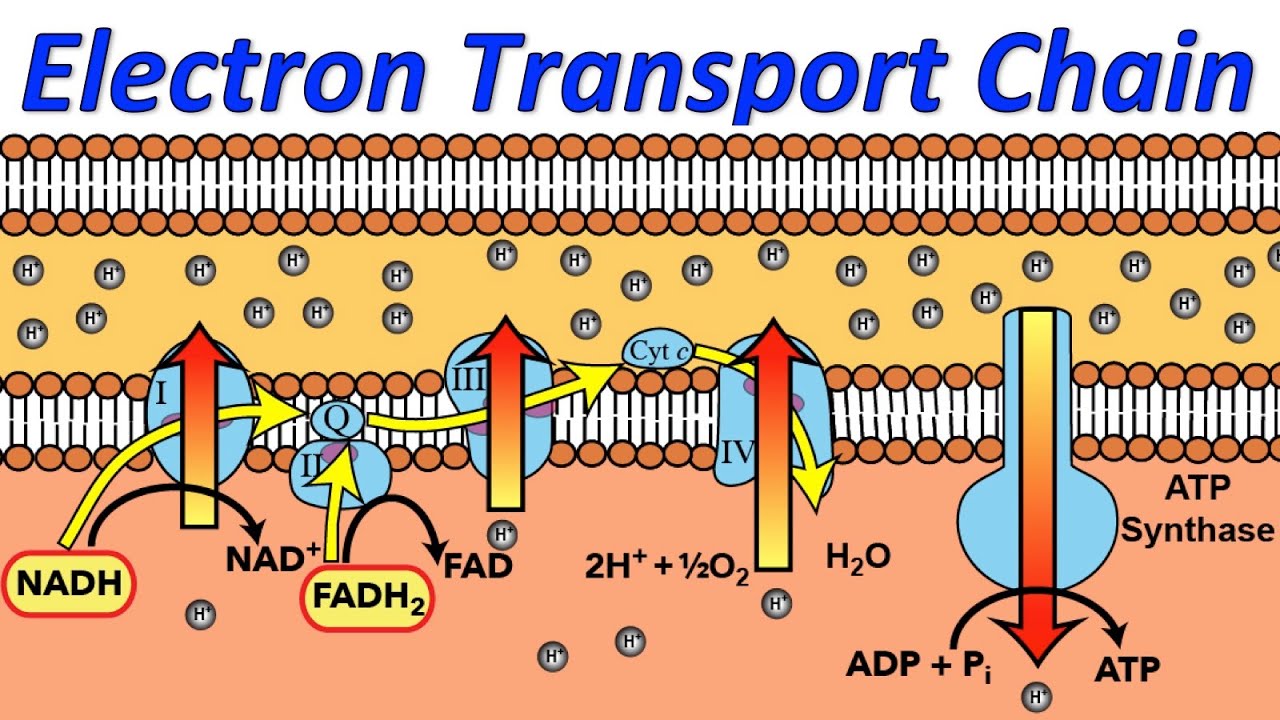
Electrons transfers down a series of electron carrier molecules // forms electron transfer chain.

- Hydrogen from glycolysis & krebs cycle combine with coenzyme NAD & FAD.
- Reduced NAD & FAD give electron from hydrogen atom they gained into the electron transfer chain
- Electrons pass through series of oxidation-reduction reactions // as electrons pass releasing energy // actively transport protons into inter-membranal space.
-  Before they are transported back into the inter-mitochodrial matrix by ATP synthase channels, protons accumulate in the inter-membranal space.
- End of the chain // protons and electrons combine with oxygen to form water // oxygen is the final acceptor of electrons in the electron transfer chain.


Releasing energy in stages

Image result for electron transfer chain


Alternative respiratory susbstrates:

Sugars and lipids and proteins can be oxidised // used a respiratory substrate without being converted into carbohydrates.


Respiration of lipids

Lipids are hydrolysed to glycerol and fatty acids.
Glycerol is phosphorylation and converted into triose phosphate // enters glycolysis pathway into 2-carbon fragments // converted to acetyl coenzyme A // enters the Kreb Cycle.
Oxidation of lipids produces 2-carbon fragment of carbohydrates and many hydrogen atoms.

Hydrogen atoms produce ATP during oxidative phosphorylation // lipids release more than double the energy for the same mass of carbohydrates.


Respiration of protein

Protein source of energy // hydrolysed to amino acids // amino group removed (deamination)
Enter the respiratory pathway at different points // 3-carbon compounds converted into pyruvate // 4- and 5-carbon compounds are converted to intermediates in the Krebs cycle.


Sunday, 14 May 2017

Energy Transfer In And Between Organisms: - Link Reaction And Krebs Cycle.

- Link reaction and Kreb cycle - 

Link reaction: 
Image result for link reaction
Pyruvate is oxidised to acetate // pyruvate loses a CO₂ and (X2) H // H is accepted by NAD to reduced NAD later used to produce ATP. 
2-Carbon acetate combines coenzyme A (CoA) = acetylcoenzyme A 


Krebs cycle


Image result for Krebs cycle

2-Carbon acetylcoenzyme A from link reaction combined with the 4-carbon molecule to produce a 6-carbon molecule
6-Carbon molecule loses carbon dioxide and hydrogen to give 4-Carbon molecules and single molecule of ATP produced as result of substrate-level phosphorylation 
4-Carbon molecule combines with new molecule of acetylcoenzyme A // cycle starts again. 


Pyruvate, link reaction and krebs cycle produces 

- Reduced coenzymes NAD and FAD // by oxidative phosphorylation potential to provide energy to produce ATP 
- 1 ATP molecule 
- 3 CO₂ molecules


2 Pyruvate molecules produced for each original glucose molecule. 

Coenzymes = molecules that enzymes require to function, includes:

NAD - important throughout repsiration 
FAD - important in the Kreb cycle 
NADP - important in photosynthesis

Respiration NAD important carrier // works with dehydrogenase enzymes catalyse removal hydrogen atoms from substrates transfer them to the other molecules involved in oxidative phosphorylation. 


Kreb cycle is important for: 

Breaking down pyruvate into CO₂ 
Producing hydrogen atoms carried by NAD to electron transfer chain prodivde energy for oxidative phosphorylation = ATP produced 
Regenerates 4 carbon molecule combines with acetylcoenzyme A 
Source of intermediate compounds used by cells - fatty acids, amino acids and chlorophyll. 


Energy Transfer In And Between Organisms: - Glycolysis

- Glycolysis -
2 Different forms of cellular respiration =

Aerobic respiration = requires O₂ produces CO₂, H₂O, ATP 

Anaerobic respiration = takes place absence of O₂ produces lactate or ethanol and CO₂ // little ATP. 

Aerobic respiration summeriesed into 4 stages 

  1. Glycolysis 
  2. Link reaction 
  3. Kreb cycle 
  4. Oxidative phosphorylation 
Glycolysis 

Image result for glycolysis a level1. Phosphorylation of glucose to glucose phosphate = glucose is made reactive with the addition of 2 phosphate molecules (phosphorylation) // Phosphate molecules come from hydrolysis of ATP (X2) ⇒ ADP. // Energy provided to activate glucose lowering activation energy for enzyme controlled reactions 

2. Phosphorylated glucose is split = Glucose is split into 3-carbon triose phosphate (X2) 

3. Triose phosphate is oxidised = H₂ removed from triose phosphate (X2) transformed into hydrogen-carrier molecule NAD to form reduced NAD

4. Production of ATP = enzyme controlled reaction converts triose phosphate into 3-carbon molecule Pyruvate // (X2) molecules of ATP are regenerated from ADP.


From ONE molecule of glucose =

2 ATP molecules // was 4 but 2 was used in initial phosphorylation of glucose - net increase of 2 molecules. 
2 reduced NAD molecules 
2 Pyruvate molecules 


Energy Transfer In And Between Organisms: - Light Independent Reaction

- Light Independent Reaction -
The Calvin Cycle - 

The products of the light dependent reaction is ATP and reduced NADP // both used to reduce glycerate-3-phosphate. 
This would have occur with or without light


Image result for the calvin cycle


1. The CO₂ diffuses into the leaf through the stomata // dissolves in water around the walls of mesophyll cells // diffuses through the cytoplasm and chloroplast membrane into the stomata of chloroplast. 
2. In stroma CO₂ reacts with 5 carbon compound   // catalysed by enzyme ribulous bisphosphate carboxylase (Rubisco)
3. Reaction CO₂ + RuBP = 2X 3-carbon glycerate 3-phosphate.
4. Reduced NADP from light dependent reaction, reduces glycerate 3-phosphatetriose phosphate using energy supplied by ATP. 
5. NADP is re-formed // goes back into the light dependent reaction = gaining protons
6. Triose phosphate converted into organic substances // starch, cellulose, lipids, glucose, amino acids, and nucleotides
7. Most triose phophate is used to generate ribulose bisphosphate // ATP is used from Light Dependent Reaction . 


Site of the light-independent reaction -

Chloroplast is adapted in carrying out the light independent reaction of photosynthesis. 

The stroma contains a fluid with all the enzymes needed for the light independent reaction. Stromal fluid is membrane-bound in the chloroplast

The stroma fluid surrounds the grana // products of light dependent stage in grana can diffuse into stroma 

Contains DNA and ribosomes // can quickly and easily manufacture some of the proteins involved in the light-independent reaction. 


Energy Transfer In And Between Organisms: - Light Dependent Reaction

- Light Dependent Reaction -

Oxidation and Reduction 

Oxidation is when the substance gains oxygen or loses hydrogen

Reduction is when the substance loses oxygen or gains hydrogen.

Oxidation results in energy being given out, whearas reduction results in it being taken in

Making of a ATP

Photoionisation is a result of the chlorophyll molecule becoming ionised

The electrons leave the chlorophyll are taken up by a molecule electron carrier // the chlorophyll is oxidised // electron carrier is reduced. 

Electrons are passed along a series of oxidation-reduction chain reactions. // A transfer chain that is located in membrane of the thylakoids

With each carrier the energy level is lowered in the electron, the energy released is used to combine the inorganic phosphate and the ADP molecule to create ATP


Chemiosmotic theory
This explains the ATP production 

    Image result for light dependent reactions
  1.  Proton pumps // protein carrier are used to pump the protons (H+) from the stroma in the thylakoid membrane. 
  2. Electrons released from photolysis drives the process as it produces protons // further increase of concentration in the thylakoid space.
  3. A concentration gradient is maintained // higher concentration = in the thylakoid space, lower concentration = in the stroma 
  4. Protons can only cross the thylakoid membrane through ATP synthesised channel proteins
  5. The channels for small granules on the membrane surface = stalked granules
  6. Protons pass through these ATP synthase channels they can cause changes to the structure of the enzyme // catalyse combination of ADP with inorganic phosphate to form ATP


Photolysis of water 

2H₂O → 4H+ + 4e⁻  + O² 

Image result for photolysis
Protons are passed out of the thylakoid space through ATP synthase channels taken up by electron carrier = NADP.
On taking up the protons the NADP reduced // the reduced NADP = main product of the light dependent stage and enters the light independent reaction.

Reduced NADP is important because it is further potential source of chemical energy to the plant.
Oxygen is a by-product from the photolysis of water is either used in respiration or diffuses out of the stomata as waste product of photosynthesis.


Site of the light-dependent reaction: 
Chloroplasts are structurally adapted to their functions of capturing sunlight. 

Thylakoid membranes have a large surface area for attachment of chlorophyll, electron carriers and enzymes carry out light-dependent reaction

Network of proteins in grana holds the chlorophyll // precise to allows maximum absorption
Granal membranes have ATP synthase channels which catalyse the production of ATP // also selectively permeable which allows establishment of proton gradient. 

The chloroplast contain both DNA and ribosomes can quickly and manufacture some of the proteins involved in light-dependent reactions.  


Energy Transfer In And Between Organisms: - Photosynthesis

- Photosynthesis - 


Site of Photosynthesis. 

Within eukaryotic plants this is the main site of photosynthesis // Within the chloroplast of cellular organelles. 

Structure of the leaf.


Image result for Structure of the leaf

  1. Large surface area = more sunlight is absorbed
  2. Arrangement of leaves = avoids overlapping // leaves do not overshadow each other. 
  3. Thin // diffusion distance is kept short = light absorbed within few micrometers 
  4. Transparent cuticle and epidermis let light through to photosynthetic mesophyll cells 
  5. Long, narrow upper mesophyll cells packed with chloroplast = collecting sunlight. 
  6. Numerous stomata = gas exchnage // short diffusion pathway from the mesophyll cells 
  7. Stomata open and closes = result of light intensity 
  8. Many air spaces in lower mesophyll layer to allow rapid diffusion in the gas phase of carbon dioxide and oxygen. 
  9. Network of xylem = brings water to leaf cells // Phloem = carries away the sugar (product of photosynthesis) 

There are 3 stages in photosynthesis. 

- Capturing the light energy 

- Light-dependent reaction 

- Light Independent reaction 


Structure of chloroplast and its role.


Image result for grana in plantThe grana = 100 discs-like structure = thylakoids // light dependent stage takes place here. 

The stroma = fluid filled matrix // light independent stage occurs here // number of other structures, for example, starch grains


Tuesday, 2 May 2017

Organisms Respond To Changes In Their Environment: - The Role Of Hormones In Osmoregulation

- Regulation of the water potential of the blood -


Osmoreceptors in the hypothalamus monitors the water potential in the blood.
If the water potential decreases in the blood, water would move out of the osmoreceptor cells by osmosis // cells decrease in volume as a result of this // signal is sent to other cells in the hypothalamus // signal then sent to the posterior pituitary gland. - this secretes the antidiuretic hormone (ADH) into the blood.

ADH makes the walls of the distal convoluted tubule and collecting duct more permeable to water.
More water is reabsorbed from these tubules into the medulla and into the blood by osmosis // small concentration of urine is produced = reduction of water loss

Specific protein receptors on cell surface membrane of these cells binds to ADH molecules // activation of phosphorylase (enzyme) within cell // vesicles within the cell move and fuse with the cell surface membrane.

Vesicles contain plasma membrane and have numerous water channel proteins (aquaporins) so when they fuse with the membrane the number of water channels is considerably increased - making cell-surface membrane more permeable to water

ADH increases permeability of collecting ducts to urea // passes out // lowering water potential of fluid around the duct.

Combining effect is more water leaves the collecting duct urea // osmosis // down a concentration gradient // re-enters the blood.

Reabsorbed water came from the blood will prevent the water potential lowering // Osmorecpetors send nerve impulses to the thirst centre of the brain encouraging the individual to seek out and drink more water.
Hypothalamus detects the rise in water potential and send fewer impulse to the pituitary gland
Pituitary gland reduces the release of ADH // permeability of the ducts return to its former state // Homeostasis and Negative Feedback.

A fall in the solute concentration of blood raises its water potential. Due to either:
1) Large volumes of water being consumed.
2) Salts used in metabolic or excreted not being replaced in the diet.


The body responds to rise in water potential:
Osmoreceptors in hypothalamus detects rise in water potential // increase frequency of the of nerve impulses to the pituitary gland to reduce its release of ADH

Less ADH // via blood // decrease in permeability of the collecting duct to water and urea

Less water is re-absorbed into the blood from the collecting ductmore dilute urine = water potential falls in the blood.

In the blood the water potential has returned to normal // osmoreceptors cause the pituitary gland to raise ADH // the levels go back to normal Negative Feedback.


Image result for - Regulation of the water potential of the blood -


Sunday, 30 April 2017

Organisms Respond To Changes In Their Environment: - Role Of The Nephron In Osmoregulation

- Nephron filtering the blood -


  1. Blood from the renal artery enters smaller arterioles in the cortex of the kidney.
  2. Arterioles splits into a glomerulus // bundle of capillaries looped inside a Bowman's capsule // Ultrafiltration takes place.
  3. Efferent arterioles that takes blood into each glomerulus // afferent arteriole take the filtered blood away from the glomerulus
  4. Efferent arterioles are smaller in diameter than afferent arterioles // blood is under high pressure. 
  5. High pressure forces liquid and small molecules in the blood out of the capillary and into the Bowman's capsule
  6. Liquid and small molecules pass through 3 layers get into Bowman's capsule enter the nephron tubules - capillary wall // basement membrane and epithelium of Bowman's capsule.
  7. Larger molecule - protein and blood cells can't pass through - stay in blood. Substances enter Bowman's capsule // glomerular filtrate 
  8. Glomerular filtrate passes along the rest of nephron - useful substances are reabsorbed along the way
  9. Filtrate flow through the collecting duct and passes out of the kidney along the ureter


Re-absorption of glucose and water by the proximal convoluted tubule
The proximal convoluted tubule is adapted to reabsorb substances: 
Its adaptations include:
- micovilli = large surface area to reasorb substances from filtrate 
- infolding of bases = large surface area to transfer reabsorbed substances back into the blood capillaries 
- high density of mitochondira to provide ATP // active transport

Process of re-absorbtion 

  1. Sodium ions actively transported out of the cells lining proximal convoluted tubule into blood capillaries // sodium concentration drops 
  2. Sodium ions diffuse down a concentration gradient from lumen of proximal convoluted tubule into epithelial lining cells // only through carrier proteins // facilitated diffusion. 
  3. Carrier proteins are specific types to carrier other specific molecules// co-transport // molecules which have been co-transported into the cell of proximal convoluted tubule then diffuse into the blood // All glucose and other valuable minerals are reabsorbed 

Maintenance of gradient of sodium ions by the loop of Henle 

Loop of Henle = responsible for water being reabsorbed from the collecting duct // concentrating the urine so it has lower water potential than blood. 

This has 2 regions 
  1. Descending limb - narrow - thin walls that are highly permeable to water. 
  2. Ascending limb - wider - thick walls that impermeable to water.

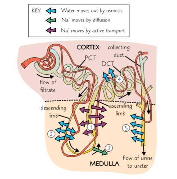
  1. Na+ ions are pumped out into the medulla using active transport // ascending limb impermeable therefore water does not move out lowering the water potential in the medulla, because there's a high concentration of ions
  2. Due to there being a lower water potential in the medulla than in descending limb, water moves out the descending limb, into medulla by osmosis // filtrate is more concentrated. The water in the medulla is reabsorbed into the blood through the capillary network
  3. Bottom of the ascending limb // Na+ ions diffuse out into the medulla, lowering the water potential further in the medulla. The ascending limb is impermeable to water, so it stays in the tubule. 
  4. Water moves out of distal convoluted tubule by osmosis and is reabsorbed into the blood.
  5. First three stages massively increase the ion concentration in the medulla, lowers the water potential. Water then moves out of the collecting duct by osmosis. The water in the medulla is reabsorbed into the blood through the capillary network


Organisms Respond To Changes In Their Environment: - Control of Blood Water Potential


- Structure Of The Nephron - 


Osmoregulation = the homeostatic control of the water potential of the blood

Structure of the mammalian kidney

In the mammal there are two kidneys found at the back of the abdominal cavity
A section through the kidney 

Image result for structure of mammalian kidney showing the position of two of the million or more nephrons in each kidney

Fibrous Capsule = outer membrane that protects the kidney 

Cortex = lighter coloured outer region made up of renal (Bowman's) capsule, convoluted tubles and blood vessels. 

Medulla = darker coloured inner region made up of loops of Henle, collecting ducts and blood vessels 

Renal pelvis = funnel-shaped cavity that collects urine into the ureter 

Ureter = tube that carries urine to the bladder 

Rental artery = supplies the kidney with blood from the heart via the aorta

Renal vein = returns blood to the heart via the vena cava 


Structure of the nephrons 

Renal (Bowman's) capsule = closed end at the start of the nephron. It cup-shaped and surrounds a mass of blood capillaries - Glomerulus. The inner layer of the renal capsule made of specialised cells - podocytes 

Proximal convoluted tubules = series of loops surrounded by blood capillaries. It's walls made epithelial cells - have microvilli 

Loop of Henle = long hairpin loop extends from cortex into medulla of kidney and back again. Surrounded by blood capillaries 

Distal convoluted tubule = series of loops surrounded by blood capillaries. Walls are made of epithelial cell - surrounded by fewer capillaries than proximal tubule

Collecting duct = tube number of distal convoluted tubules from a number of nephrons empty. Lined by epithelial cells and becomes increasingly wide as it empties into the pelvis of kidney


Associated with each nephron there are number of blood vessels:

Afferent arteriole = tiny vessel arise from renal artery - supplies the nephron with blood // Afferent arteriole enters the renal capsule of the nephron 

Glomerulus = many-branched knots of capillaries from which fluid is forced out of the blood. Glomerulus capillaries recombine.

Efferent arterioles = tiny vessel leaves the renal capsule // smaller diameter than afferent arteriole causes an increase in blood pressure within the glomerulus. Efferent arterioles carries blood away from the renal capsule

Blood capillaries = concentrated network of capillaries surrounding the proximal convoluted tublue, loop of Henle mineral salts, glucose, and water. These capillaries merge to form venules, merging to form the renal vein


Organisms Respond To Changes In Their Environment: - Diabetes And Its Control

- Diabetes and its control - 


Types of sugar diabetes 


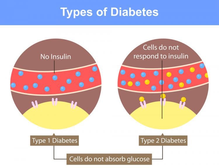
Type 1 - insulin dependent // body unable to produce insulin 

The body attacks its own cells - 𝛽 cells - result of autoimmune response. 
This develops quickly over a few weeks once the individual is born. Usually develops at a very young age. 

Type 2 - insulin independent // body glycoproteins receptors have lost or losing their responsiveness to insulin // body producing an insufficient amount of insulin
This develops in people over age of 40 - increases due to obesity and poor diet
Symptoms are less severe and may go unnoticed
90% of people have type II diabetes 


Control of diabetes 

Diabetes can be successfully treated which depends on the type of diabetes 

Type 1 diabetes = controlled by injections of insulin - cannot be taken orally as it is an enzyme and would be digested. Typcially taken 3-4 times a day. Dose of insulin would match the dose intake of glucose // glucose concentration is monitored using biosensors

Type 2 diabetes = controlled by regulating the intake of carbohydrates in the diet // exercise is also taken into account. Supplemented by injections of insulin by the use of drugs that stimulate insulin production


Image result for insulin type 1 and type 2


Organisms Respond To Changes In Their Environments: - Hormones And The Regulation Of Blood Glucose Concentration

- Hormones and their mode of action - 

Hormones differ chemically but there are characteristics which would be the same also. 

Hormones :

- Produced in glands // secrete hormones directly into blood
- Carried in blood plasma to cells which are acted on // target cells // specific receptors on their cell-surface membrane // complementary to specific hormones.
- Effective in very low concentrations // widespread // long-lasting.


 Second Messenger Model 

2 hormones are used - Adrenaline and glucagon 

  1. Adrealine binds to the transmembrane protein receptor within the cell-surface membrane of the liver cell. 
  2. Binding of the adrealine causes the protein to change the shape on the inside of the membrane
  3. Change in shape of the protein leading to the activation of an enzyme - adenly cyclase // activated adenly cyclase converts ATP to cyclic AMP (cAMP) 
  4. cAMP acts as second messenger that binds to the protein kinase enzyme, its activiated by changing its shape.
  5. Active protein kinase enzyme catalyses the conversion of glycogen to glucose which moves out of the liver cell // facilitated diffusion // in to the blood through channel proteins
Image result for second messenger model

Role of the Pancrease in regulating blood glucose 

It produces enzymes - 
Protease, amylase and lipase for digestion and hormones
Insulin and glucagon for regulating blood glucose concentration

There are groups of hormone-producing cells - islets of Lnagerhans - cells of the islets of Langerhans:
- ɑ cells, larger and produce the hormone glucagon
- 𝛽 cells, smaller and produce the hormone insulin 

Role of the liver in regulating blood sugar 

There are three important processes involved with regulating blood sugar which takes place in the liver:

Glycogenesis - this is the conversion of glucose into glycogen
This occurs when the blood glucose concentration is higher than normal. The liver removes the glucose from the blood and converts it into glycogen

Glycogenolysis - this is the breakdown of glucagon into glucose.
This occurs when the blood glucose concentration is lower than normal. The liver converts glycogen into glucose which is then diffused into the blood

Gluconeogenesis - this is production of glucose from sources other than carbohydrates
This occurs when glycogen supply is exhausted. The liver produces glucose from non-carbohydrate sources - amino acids and glycerol


Regulation of blood glucose concentration 


Factors that influence blood glucose concentration
  1. Directly from the diet = glucose is absorbed from the hydrolysis of carbohydrates 
  2. From the hydrolysis of glycagon in the small intestine = glycogenolysis // stored in liver and muscle cells 
  3. From gluconeogenesis 


Insulin and the 𝛽 cells of the pancreas 
when combined with receptors insulin brings about:

- Change in tertiary strucute of the glucose transport carrier proteins - changing shape and open - allows more glucose into the cells by facilitated diffusion.
- Increase in number of carrier proteins responsible for glucose transport in the cell-surface membrane. Protein from which these channels are made is part of the vesicles. // rise in insulin concentration the vesicles fusing with the cell-surface membrane increasing the number of glucose transport channels. 
- Activation of enzyme that convert glucose to glycogen and fat

Blood glucose is lowers through the following ways:

- Increasing the rate of absorption of glucose into the cells - muscle cells
- Increasing rate of respiration of the cells // uses more glucose // increasing the uptake from the blood
- Increasing glycogenesis in liver and muscle cells
- Increasing the rate of conversion of glucose to fat

The processes taking place = Negative feedback // removal of glucose reduced the secretion of insulin from 𝛽 cells


Glucagon and the ɑ cells of the pancreas 
ɑ cells detect the fall in blood glucose concentrations releasing the hormone glycogen directly into the blood plasma
Glycogen action include: 
- Attaching to specific protein receptors on cell-surface membrane of liver cells 
- Activating enzymes convert glycogen to glucose 
- Activating enzymes involved in conversion of amino acids and glycerol into glucose = glyconeogenesis 
This returns the blood glucose concentration to the optimum

Image result for alpha and beta cells in the pancreas

Role of adrenaline in regulating the blood glucose level 

Adrenaline raises the blood glucose concentration by:
- Attaching to protein receptors on the cell-surface membrane of target cells 
- Activating enzymes that causes the breakdown of glycogen to glucose in the liver

Adrenaline is produced when there is a period of excitement or stress

Hormone interaction in regulating the blood glucose

Insulin and glycagon act in opposite directions = antagonistically 
These two hormones are sensitive to control of the blood glucose concentration // blood glucose concentration is not constant there are fluctuations around the optimum temperature

When writing about negative feedback its important to mention that the secretion of a hormone - insulin - would result in the reduction of its own secretion


Organisms Respond To Changes In Their Environments: - Feedback Mechanisms

- The control mechanisms -

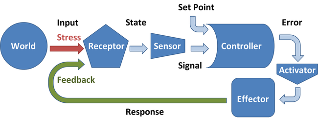
Control of any self regulating system involves a series of stages

- Optimum point
- Receptor
- Coordination
- Effector
- Feedback mechanism

Homeostatic systems detect change and respond by negative feedback.

1) The receptorcommunication system and effectors is the foundation of the homeostatic system
2) Receptors are there to detect when there is a change in temperature - this would be when temperature is too high or too low. Via the nervous system or hormonal system the information is passed on to the effector. 
3) The effectors are suppose to counteract the change // bring the level back to an optimum
4) Negative feedback = process of bring the level to an optimum temperature
5) Negative feedback keeps the body's temperature at an optimum which is above 0.5℃ or below 37℃.
6) However if they change is too drastic then the Negative feedback would be unable to counteract the change. This can be when there has been prolonged exposure to cold weather


There are multiple Negative feedback mechanisms ⇒ more control. By only having one negative feedback system ⇒ there is a slower response and less control

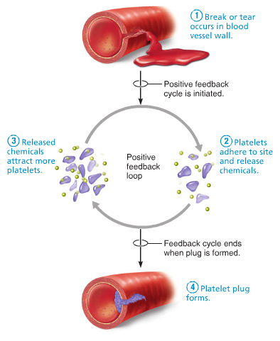
Positive feedback mechanisms can amplify a change from the normal level
1) some changes can trigger a positive feedback // amplifying the change. 
2) Increasing the level further away from the normal level.
3) Positive feedback rapidly activates something // Blood clot after a deep cut. 
4) Positive feedback can occur when homeostatic system breaks down e.g. Being too cold for too long.

Positive feedback is not involved in homeostasis because it does not keep the internal environment stable.


Image result for positive feedback homeostasis

- Coordination of control mechanisms -

Control system normally have receptors and effectors allows them to have separate mechanisms that produce a positive movement towards an optimum. Allows greater degree of control of the particular factor being regulated. 


Thursday, 27 April 2017

Organisms Respond To Changes In Their Environments: - Principles Of Homeostasis

- Principles Of Homeostasis -

What is Homeostasis?

This is the maintenance of an interval environment within restricted limits in organisms.
This is to ensure that the chemical make-up, volume and other features of blood and tissue fluid within restricted limits are maintained.

Homeostasis is the maintenance of a stable internal environment.
Any changes that are made to the external environment can affect the internal environment of an organism.

To prevent this, homeostasis is involved in control systems that would maintain the constant internal environment.
This allows virtual cells to function normally and prevent damage to them and they are kept in a stable environment.
Factors that would be maintained ⇒ the core body temperature and blood pH.

Image result for Homeostasis

This is because both pH and temperature are factors that effect enzyme activity and enzymes control the rate of metabolic reactions.

Temperature: if it is too high or at an extreme the enzymes would become denatured.
This is due to the hydrogen bonds breaking that holds them in their 3D shape. Thus changing the active site, which leads to an ineffective catalyst.
If the temperature is too low the enzyme activity is reduced and it slows down the rate of metabolic reactions.

In order for the rate of enzyme activity to remain at a constant the enzymes have an optimum temperature at which they work best at. In humans, this would be 37oC.

pH: if the pH is too high or too low // at either ends of extremes// the enzymes denature. Hydrogen bonds that hold the shape of the enzymes are broken. Therefore, the active site of the enzyme is changed which makes it ineffective catalyst. The metabolic reactions are less efficient.
The highest rate of enzyme activity remains at an optimum pH // usually remains around pH 7.
But this varies for some enzymes // enzymes for the stomach work best at a low pH.
Because cells need glucose for energy it is important to maintain the right concentration of glucose in the blood.

Image result for Homeostasis blood pH
Blood glucose concentration also affects the water potential of blood - there is the potential that water molecules diffuse in and out of or into a solution.
If blood glucose concentration is high ⇒ water potential of blood is reduced // water molecules would then diffuse out of the cell // osmosis // cell will shrivel up and die.

If blood glucose concentration is too low ⇒ cells cannot carry out normal activities there isn't enough glucose for respiration to provide energy.


- The control mechanisms -

Control of any self regulating system involves a series of stages

- Optimum point
- Receptor
- Coordination
- Effector
- Feedback mechanism

Homeostatic systems detect change and respond by negative feedback.

1) The receptor, communication system and effectors is the foundation of the homeostatic system
2) Receptors are there to detect when there is a change in temperature - this would be when temperature is too high or too low. Via the nervous system or hormonal system the information is passed on to the effector. 
3) The effectors are suppose to counteract the change // bring the level back to an optimum
4) Negative feedback = process of bring the level to an optimum temperature
5) Negative feedback keeps the body's temperature at an optimum which is above 0.5℃ or below 37℃.
6) However if they change is too drastic then the Negative feedback would be unable to counteract the change. This can be when there has been prolonged exposure to cold weather


There are multiple Negative feedback mechanisms ⇒ more control. By only having one negative feedback system ⇒ there is a slower response and less control

Positive feedback mechanisms can amplify a change from the normal level
1) some changes can trigger a positive feedback // amplifying the change. 
2) Increasing the level further away from the normal level.
3) Positive feedback rapidly activates something // Blood clot after a deep cut. 
4) Positive feedback can occur when homeostatic system breaks down e.g. Being too cold for too long.

Positive feedback is not involved in homeostasis because it does not keep the internal environment stable.


Image result for positive feedback homeostasis

- Coordination of control mechanisms -

Control system normally have receptors and effectors allows them to have separate mechanisms that produce a positive movement towards an optimum. Allows greater degree of control of the particular factor being regulated. 


Saturday, 22 April 2017

Organisms Respond To Changes In Their Environment: - Control Of Heart Rate.

- Control Of Heart Rate -

The Autonomic Nervous system

It has two subdivisions:

  1. Sympathetic nervous system - stimulates effectors so speeds up any activity. Acts rather like an emergency controller // allows us to cope with stressful situations heightening our awareness and prepare us for the activity. 
  2. Parasympathetic nervous system -  inhibits effectors so it slows down any activity. It controls activities to a normal resting conditions // conserves the energy and the body's reserves are replenished. 
The system oppose each other - antagonistic. If one contracts a muscle the other relaxes a muscle. Each doing the opposing job 

- Control Of Heart Rate -
Cardiac muscle = myogenic // able to contract without receiving signals.

  1. Sinoatrial node (SAN) is in the wall of the right atrium, recieves an electrical activity which spreads across both atria casuing them to contract. SAN is like a pacemaker, setting the rhythm of the heart. 
  2. Layer of non-conductive collagen tissue // atrioventricular septum // stops waves of electrical activity from being passed directly to the ventricles
  3. The waves are then passed to/enters the atrioventricular nodes (AVN) This is in between the atria 
  4. There is a slight delay when the AVN passes the electrical activity to the bundle of His // a series of muscle fibres (Purkyne tissue) which collectively make up the bundle of His // the atria would have had to emptied before the AVN reacts
  5. Bundle of His grouped of muscle fibres responsible for conducting the waves of electrical activity between ventricles to apex. // bottom of the heart. 
  6. Purkyne tissue carries the waves of electrical activity into a muscular walls of both ventricles, both contracting at the same time // from the bottom up


Image result for SAN and AVN


- Modifying the resting heart rate - 

Adult human hearts has a resting heart beat of 70BPM // important to be able to change this heart rate in order to meet the varying demands for oxygen.
During exercise the heart rate is likely to increase to double the original resting heart rate.

Medulla Oblongata = controls the changes to the heart rate in the a region of the brain // sub-divided into two centres:
  1. Centre that increases heart rate - linked to sinoatrial node - sympathetic nervous system (SNS)
  2. Centre that decreases heart rate - linked to sinoatrial node - parasympathetic nervous system (PNS) 
- Control by chemoreceptors -

Pressure receptors = baroreceptors in the aorta & carotid arteries // stimulated by high & low blood pressure

Chemical receptors = chemoreceptors in the aorta, carotid arteries, and medulla // monitor oxygen levels in the blood and carbon dioxide and pH - both indicators of O2 levels

This works by: 

  1. Blood has a higher than normal concentration of carbon dioxide // pH is lowered
  2. Chemorecpetors in walls of carotid arteries // aorta detect & increases the frequency of nerve impulses to the centre in medulla oblongata - increases heart rate
  3. This centre increases the frequency of impulses via SNS to SAN increasing the rate of production of electrical waves by SAN thus increasing the heart rate
  4. Increased blood flow that causes leads to lungs removing the carbon dioxide, CO2 concentration return to normal in the blood
  5. pH of the blood rises to normal // the chemoreceptors in the walls of the carotid arteries & aorta reduce frequency of nerve impulse to the medulla oblongata 
  6. Medulla oblongata reduces frequency of impulses to SAN, leads to a reduction in the heart rate. 


Image result for control of heart by chemoreceptors


- Control by pressure receptors - 


The pressure receptors = occurs within heart walls // carotids arteries & aorta // sequence of that follows changes in activity levels:


  • When blood pressure is higher than normal = receptors transmits more nervous impulses to the centre in medulla oblongata = decreases heart rate. Impulses sent via PNS (Parasympathetic) to SAN from centre
  • When blood pressure is lower than normal = receptors transmit more nerve impulses to centre in medulla oblongata = increases heart rate. Impulses sent via SNS (Sympathetic) to SAN from centre

Biological Molecules: - Carbohydrates

- Carbohydrates -   - Monosaccharides -  The monomer for carbohydrates is called monosaccharides.  Common for...