Thursday 27 April 2017

Organisms Respond To Changes In Their Environments: - Principles Of Homeostasis

- Principles Of Homeostasis -

What is Homeostasis?

This is the maintenance of an interval environment within restricted limits in organisms.
This is to ensure that the chemical make-up, volume and other features of blood and tissue fluid within restricted limits are maintained.

Homeostasis is the maintenance of a stable internal environment.
Any changes that are made to the external environment can affect the internal environment of an organism.

To prevent this, homeostasis is involved in control systems that would maintain the constant internal environment.
This allows virtual cells to function normally and prevent damage to them and they are kept in a stable environment.
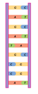
Factors that would be maintained ⇒ the core body temperature and blood pH.

Image result for Homeostasis

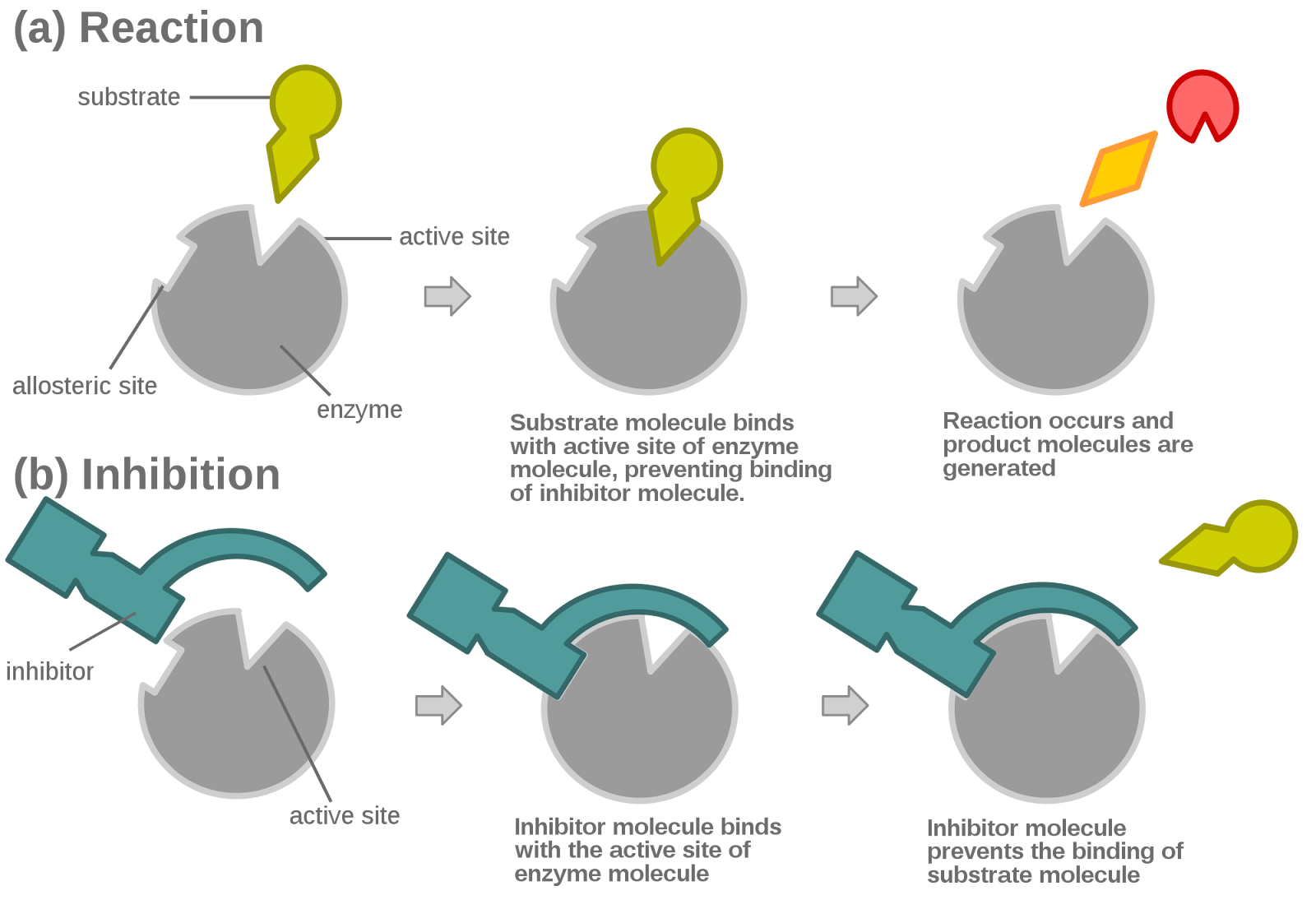
This is because both pH and temperature are factors that effect enzyme activity and enzymes control the rate of metabolic reactions.

Temperature: if it is too high or at an extreme the enzymes would become denatured.
This is due to the hydrogen bonds breaking that holds them in their 3D shape. Thus changing the active site, which leads to an ineffective catalyst.
If the temperature is too low the enzyme activity is reduced and it slows down the rate of metabolic reactions.

In order for the rate of enzyme activity to remain at a constant the enzymes have an optimum temperature at which they work best at. In humans, this would be 37oC.

pH: if the pH is too high or too low // at either ends of extremes// the enzymes denature. Hydrogen bonds that hold the shape of the enzymes are broken. Therefore, the active site of the enzyme is changed which makes it ineffective catalyst. The metabolic reactions are less efficient.
The highest rate of enzyme activity remains at an optimum pH // usually remains around pH 7.
But this varies for some enzymes // enzymes for the stomach work best at a low pH.
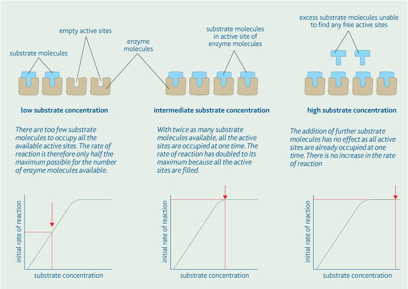
Because cells need glucose for energy it is important to maintain the right concentration of glucose in the blood.

Image result for Homeostasis blood pH
Blood glucose concentration also affects the water potential of blood - there is the potential that water molecules diffuse in and out of or into a solution.
If blood glucose concentration is high ⇒ water potential of blood is reduced // water molecules would then diffuse out of the cell // osmosis // cell will shrivel up and die.

If blood glucose concentration is too low ⇒ cells cannot carry out normal activities there isn't enough glucose for respiration to provide energy.


- The control mechanisms -

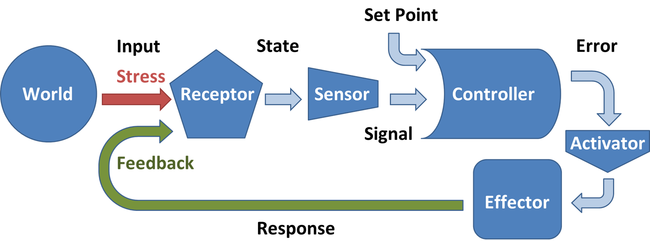
Control of any self regulating system involves a series of stages

- Optimum point
- Receptor
- Coordination
- Effector
- Feedback mechanism

Homeostatic systems detect change and respond by negative feedback.

1) The receptor, communication system and effectors is the foundation of the homeostatic system
2) Receptors are there to detect when there is a change in temperature - this would be when temperature is too high or too low. Via the nervous system or hormonal system the information is passed on to the effector. 
3) The effectors are suppose to counteract the change // bring the level back to an optimum
4) Negative feedback = process of bring the level to an optimum temperature
5) Negative feedback keeps the body's temperature at an optimum which is above 0.5℃ or below 37℃.
6) However if they change is too drastic then the Negative feedback would be unable to counteract the change. This can be when there has been prolonged exposure to cold weather


There are multiple Negative feedback mechanisms ⇒ more control. By only having one negative feedback system ⇒ there is a slower response and less control

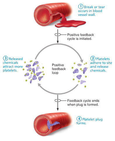
Positive feedback mechanisms can amplify a change from the normal level
1) some changes can trigger a positive feedback // amplifying the change. 
2) Increasing the level further away from the normal level.
3) Positive feedback rapidly activates something // Blood clot after a deep cut. 
4) Positive feedback can occur when homeostatic system breaks down e.g. Being too cold for too long.

Positive feedback is not involved in homeostasis because it does not keep the internal environment stable.


Image result for positive feedback homeostasis

- Coordination of control mechanisms -

Control system normally have receptors and effectors allows them to have separate mechanisms that produce a positive movement towards an optimum. Allows greater degree of control of the particular factor being regulated. 


Sunday 23 April 2017

Nucleic Acids: - DNA Replicationn

- DNA Replication - 
DNA replication: cell division occurs in 2 main stages:
  • Nuclear division: nucleus divides
  • Cytokinesis - the whole cell divides
Semi-conservative replication ~ this ensures the genetic continuity between generations.

The enzyme DNA helicase breaks the hydrogen bonds between complementary bases in polynucleotides strands, causing the double helix to unwind/unravel.


.
The enzyme DNA polymerase joins adjacent nucleotide's in a condensation reaction.


The exposed nucleotide's act as a template to which the complementary free nucleotide's bid by specific base pairing


2 identical molecules of DNA are formed. This is due to each molecule retaining half of the original DNA materialWatson and Crick model of DNA replication suggested that the original DNA molecule splits into 2 separate strands therefore each of the new molecules would have one strand of the original material. This would be the semi-conservative model.  The conservative model suggests that the original molecule remained intact and the other molecule formed would be made of entirely new material

Image result for the conservative replication


Nucleic Acids: - Structure of RNA and DNA

- Structure of DNA and RNA -


Deoxyribonucleic acid and ribonucleic acid are important information-carrying molecules. In all living cells, DNA holds genetic information and RNA transfers genetic information from DNA to the ribosomes.
DNA molecule is a double helix with two polynucleotides chains held together by hydrogen bonds between specific complementary base pairs.
  • Adenine is complementary to thymine
  • Guanine is complementary to cytosine
Ribosomes are formed from RNA and protein.
Both DNA and RNA are polymers of nucleotides. Each nucleotides is formed from

  • Pentose sugar
  • Phosphate group
  • Nitrogen-containing organic base

Nitrogen containing bases are:
  • Adenine - A
  • Thymine - T
  • Guanine - G
  • Cytosine - C
  • Uracil - U ( RNA molecules only // replaces Thymine (T))
Pentose sugar, phosphate group and organic base are joined as result of condensation reactions, forming a mononucleotide.
2 mononucleotides may in turn be joined by the condensation reaction between the deoxyribose sugar of one mononucleotide and the phosphate group of another. The bond formed between them is called a phosphodiester bond.
Image result for structure of rna and dna

RNA structure:
  • RNA molecules is a relatively short polynucleotide chain
  • The pentose sugar is ribose and the organic bases are adenine, guanine, cytosine, and uracil.
DNA structure // Double helix structure (DNA) ~
  • The pentose sugar is deoxyribose and the organic bases are adenine, thymine, guanine and cytosine.
  • Alternating phosphate and deoxyribose molecules make up the ‘uprights’ and pairs of organic bases comprise the ‘rungs’. The complementary base pairing ensures a standard ‘rung length’.

Stability of DNA ~
The phosphodiester backbone protects the more chemically reactive organic bases.
Hydrogen bonds link the organic base pairs forming bridges between the phosphodiester uprights
The higher the proportion of G-C pairings, with 3 hydrogen bonds, A-T pairings have 2 hydrogen bonds.
Function of DNA ~
-
DNA is responsible for passing genetic information from cell to cell.

- Adaptations.

- Large molecule - carries an immense amount of genetic information.

- Base pairing leads to DNA being able to replicate and transfer into as mRNA.

- The base pairs are found in the helical cylinders therefore they are protected from outside chemical and physical forces.

- Mutations in DNA due to the stability of the molecule. // whether or not a mutation would occur.

- Separate strands are held by hydrogen bonds - easy to separate.

- The simplicity of DNA led to many scientists doubt that it carried the genetic code.


Biological Molecules: - Enzyme Inhibition

- Enzyme Inhibition - 


Enzyme inhibition: enzyme inhibitors are substances that directly or indirectly interfere with the function of the active site of an enzyme, reducing its activity.
Competitive inhibitors: inhibitors have a molecular shape which would be similar to the substrate, which allows them to occupy the active site instead of the substrate, competing with the substrate molecule.
If the substrate concentration is increased, the effect of the inhibitor rescued.
The inhibitors is not permanently bound to the active site thus when it leaves, another molecule can take it place.
The concentration of the inhibitor determines how long it will take for all substrate molecules to occupy the active site.
Image result for competitive inhibition


- Non competitive inhibitors -
They alter the shape of the enzyme and active site which means that substrate molecules can no longer occupy it and so the enzyme cannot function. Because the inhibitor and the substrate are not competing for the same site, an increase in substrate concentration does not decrease the effect of the inhibitor.


Image result for non-competitive inhibition

Biological Molecules: - Factors Affecting Enzyme Action

- Factors Affecting Enzyme Action - 

There are many factors that effect enzyme action, this includes:

  1. Temperature.  
  2. pH levels.
  3. Concentration
However before delving into detail about how they effect the enzyme action. First, we have to see how enzyme-catalysed reactions are measured. 


To measure the progress of an enzyme-catalysed reaction the time course is measured

2 changes that are frequently measured:

> Formation of the products of the reaction
> Disappearance of substrate.


Measuring rate of change 


The change in the rate of reaction can be measured at any point on the curve of a graph. 

The gradient is equal to the gradient of the tangent to the curve at the point the tangent is the point at which a straight line touches the curve but without cutting across it. 


You can draw the tangent to the curve at the point shown. Using this line you can find the gradient 

     a 
 = ㄧ     
     b

Effects of different factors on the rate of enzyme action it is important to stress the fundamental experimental technique of changing only a single variable in each experiment. 

Rate of an enzyme reaction all the other variables must be kept constant when investigating the effects of a named variable. 


Another thing to remember is that the active site and substrate is not the same as a lock and key.  

Temperature ● pH ● Concentration 
Factors effecting enzyme action. 


Image result for Factors Affecting Enzyme Action


Temperature: 

When there a temperature increase, there is an increase in kinetic energy of molecule. 
The molecules move around rapidly colliding with each other more often. 
Enzyme-catalysed reaction = enzyme and substrates molecules come together more often in a given time. 
More effective collisions = More enzyme-substrate complexes being formed

At first the substrate does not fit as easily into the changed active site = slowing rate of reaction. 
Human enzymes begin at temperature of 45℃. 
Around 60℃ enzyme is so disrupted its stops working together = the enzyme denatures
Denaturation is a permanent change and enzyme does not function again. 
Many enzymes have an optimum temperature of around 40℃ in the human body. 

Optimum temperature = the most/best favourable point or degree or condition at which an organism can work at which would obtain the best results


pH: 

pH of a solution is the measure of hydrogen ion concentration. 

The pH of a solution is calculated using : 

pH = -log₁₀[H⁺] 

H⁺ concentration of 1 x 10⁻⁹ therefore has a pH of 9 

An increase or decrease in pH reduces rate of enzyme action. If the pH level is extreme the enzyme becomes denatured. 

The pH level affects enzymes works in two ways:
  1. pH alters the charge on the amino acids that make up the active site of the enzyme = substrates can no longer attach to the active site. In other words, enzyme-substrate complex cannot be formed. 
  2. pH may cause the bonds maintaining the enzyme's tertiary structure to break = active site changes. 


Concentration: 

Image result for effect of enzyme concentration on rate of enzyme action

The active site can be reused to repeat the procedure on another substrate molecule once it is free. This means that enzymes are not used up during the chemical reactions, resulting in them working efficiently at very low concentrations.
  1. Low enzyme concentration: there are too few enzyme molecules to find an active site at one time.
  2. Intermediate enzymes concentration: all the substrate molecules can occupy an active site as the same time. The rate of reaction had doubled to it's maximum as a result.
  3. High enzyme concentration: adding more enzyme molecules has no effect as there are already enough active sites to accommodate all the substrate molecules.


Biological Molecule: - Non- Reducing Sugar Test

- Test For Non-Reducing Sugars - 


Test for non-reducing sugars: some disaccharides are reducing sugars and we can use Benedict's test to detect them. Sucrose is known as a reducing sugar because it doesn't change the colour of Benedict's reagent when heated with it.


  1. Liquify sample.
  2. Add 2cm³ of the food sample to 2cm³ of Benedict's reagent.
  3. Place the test tube in a gently boiling water bath for 5 minutes. If the Benedict's reagent does not change colour then a reducing sugar is not present.
  4. Add another 2cm³ of the food sample to 2cm³ of dilute hydrochloric acid in a test tube and place the test tube in gently boiling water bath for 5 minutes. The hydrochloric acid will hydrolyse any disaccharides present into its constituent monosaccharides.
  5. Add sodium hydrogen carbonate solution to neutralise the hydrochloric acid. Test with pH paper to check that solution is alkaline
  6. Retest the resulting solution, if non reducing sugar present the sample would turn orange-brown colour


Image result for test for reducing sugars

Biological Molecule: - Reducing Sugars Test

- Test For Reducing Sugars - 


Test for reducing sugars:
All monosaccharides and some disaccharides are reducing sugars. A reduction reaction is a chemical reaction involving the gain of electrons or hydrogen. Reducing sugars that can donate electrons to another chemical. Reducing sugars can be tested for using Benedict's reagent which is an alkaline solution of copper II sulphate. An insoluble red precipitate of copper oxide is formed when reducing sugars are heated with benedict's reagent.
Methods:

  1. Add 2cm³ of the food sample to be tested to be tested to the test tube (this should be in liquid form).
  2. Add an equal volume of Benedict's reagent.
  3. Heat the mixture in a gently boiling water for 5 minutes.

Image result for carbohydrates structure

Biological Molecules: - Test For Lipids

- Test For Lipids - 


Test for lipids:
  1. Take a completely dry and grease-free test tube.
  2. Add 5cm³ of ethanol.
  3. Shake the tube thoroughly to dissolve any lipid in the sample.
  4. Add 5cm³ of water.
  5. A cloudy - white colour indicates indicates the presence of a lipid.
  6. Repeat using water instead of the sample, the final solution should remain clear.
The cloudy colour is due to any lipid in the sample being finely dispersed in the water to form an emulsion.

Image result for test for lipids

Biological Molecules: - Tests for Starch

- Test For Starch - 

Test for starch:


  1. Place 2cm³ of sample being tested into a test tube.
  2. Add a few drops of iodine solution and shake or stir.
  3. The presence of starch is indicated by the blue-black coloration.


Image result for test for starch in food

Biological Molecules: - Carbohydrates

- Carbohydrates -   - Monosaccharides -  The monomer for carbohydrates is called monosaccharides.  Common for...